
Corneal Disease
 The cornea is the outermost layer of your eye. Covering the front surface of your eye, it’s dome-shaped and clear. Your cornea helps focus your vision, providing 65 to 75 percent of the focusing power of your eye. Even though your cornea is clear and looks simple, it contains specialized tissue that differs from most tissues in your body because it contains no blood vessels to protect it from infection. The cornea is nourished by your tears and aqueous humor, which is the fluid behind the cornea in the front part of your eye.
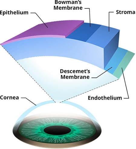
Your cornea serves as a barrier against dirt, germs, and other things that can harm your eye. It shares the protection of your eye with the sclera (the white of your eye), eye sockets and eyelids. The cornea works as a filter to keep the damaging UV rays of the sun from harming your retina. Your cornea has five layers with membranes in between. Each layer has its own important function:
- Epithelium: The cornea’s outside layer, the epithelium functions primarily as a shield to protect the eye from foreign objects like bacteria, dust and water. It’s also a smooth surface that absorbs nutrients and oxygen from your tears for the other corneal layers. The epithelium has thousands of tiny nerve endings, which is why rubbing or scratching your eye causes pain. The epithelial cells are attached to an anchor called the basement membrane.
- Bowman’s Membrane: The second layer, behind the basement membrane, Bowman’s membrane is a transparent film made up of protein fibers known as collagen. If you injure your Bowman’s membrane, it can become scarred. Depending on the size and location of the scars, this can result in vision loss.
- Stroma: The next layer down, the stroma is the thickest layer within the corneal structure. It’s made up mostly of collagen and water. The collagen helps the cornea keep its form, elasticity and strength. The stroma’s unique arrangement, shape and spacing of collagen proteins produce the light-conducting ability of your cornea.
- Descemet’s Membrane: Located below the stroma, Descemet’s membrane is made up of thin but strong tissue that creates a protective barrier against injury and infection. Composed of a different type of collagen fibers, this membrane heals well after injury.
- Endothelium: The innermost layer of your cornea, the cells of the endothelium function to keep the cornea clear by maintaining a fluid balance. If the endothelium didn’t perform this function, the stroma would become cloudy. The endothelium also adds collagen to the basement membrane. Endothelial cells that are damaged or destroyed are not repaired or replaced by your body.
Corneal Problems
Most minor injuries to your cornea heal on their own. Deeper or more serious injuries can leave scars on the cornea, which can impair your vision. If you have a disease or damage to your cornea, you may experience symptoms such as:
- Eye pain
- Light sensitivity
- Blurry or reduced vision
- Headache
- Nausea
- Fatigue
- Inflammation
- Redness
Corneal injuries should always be evaluated with a thorough consultation and examination by a physician for an accurate diagnosis and treatment plan as it may lead to scarring and possibly blindness.
Corneal Dystrophy
This condition exists when one or more parts of your cornea become cloudy because of a buildup of material on the outside layer. Corneal dystrophy:
- Usually is inherited
- Affects both eyes
- Doesn’t affect other parts of your body and isn’t related to any other diseases in your body
- Progresses naturally
- Happens in people who are otherwise healthy
Corneal dystrophy can affect your vision in a number of ways. Sometimes, it causes no impairment and is only discovered during an eye vision test. At other times, it can lead to severe vision loss. It’s also been known to cause recurring pain without any affect on your vision. There are four different types of corneal dystrophy:
- Keratoconus: The most common type of corneal dystrophy, it can occur in as many as one in every 2000 Americans, mostly teens and adults in their 20s. It’s an abnormal curvature of the cornea that can lead to increased light sensitivity, nearsightedness and astigmatism. The condition usually appears in both eyes, but can be treated with contact lenses to correct the distortions. For most people, the condition stabilizes after a few years and causes no further problems.
- Fuch’s Dystrophy: This disease, which affects both eyes, progresses quite slowly. It’s more common in women than men. Your vision gradually worsens over time, although you may not notice any issues until you are 50 or 60 years old. Fuch’s Dystrophy is caused by the slow degradation of the corneal endothelial cells. Your vision becomes blurred over time. Other symptoms include glare, distorted vision, poor night vision and halos around lights at night. Treatment involves reducing the swelling with drops or using soft contact lenses. In severe cases, your ophthalmologist may recommend a corneal transplant.
- Lattice Dystrophy: This condition gets its name from the appearance of lattice-like deposits that develop on your stroma. As more and more deposits form, they become opaque, impairing your vision. It can happen at any time during your life, but usually begins between ages two and seven. In severe cases, the deposits can converge under your epithelium, causing erosion, which can be extremely painful if the nerves that line your cornea are exposed. Your eye doctor can prescribe drops or ointments to reduce friction. You may need an eye patch to keep your eye from moving too much. It takes six to eight weeks to heal. In some cases, by the time you reach age 40, you may have so much corneal scarring that you require a corneal transplant.
- Map–Dot-Fingerprint Dystrophy: Also known as epithelial basement membrane dystrophy, this condition can occur when your basement membrane doesn’t develop normally, creating folds created in the tissue. The folds appear as grey shapes that look like continents, opaque dots or concentric lines that look like a fingerprint. Symptoms include excessive tearing, blurred vision, morning pain, light sensitivity and a feeling that there’s something in your eye. This disease affects both eyes and usually occurs in adults age 40 to 70. It’s possible for you to have this dystrophy and have no symptoms at all. Some people do feel pain. This condition can come and go, but you can manage the symptoms with drops or ointment. If this doesn’t work, there are surgical options such as corneal scraping, laser surgery or anterior corneal puncture.
If you are having any abnormal visual symptoms, you should always be evaluated with a thorough consultation and examination by a physician for an accurate diagnosis and treatment plan as it may be a symptom or sign of a serious illness or condition.
Important Reminder: This information is only intended to provide guidance, not a definitive medical advice. Please consult eye doctor about your specific condition. Only a trained, experienced board certified eye doctor can determine an accurate diagnosis and proper treatment.
Do you have any questions about Corneal Disease and Corneal Dystrophy? Would like to schedule an appointment with the best ophthalmologist in NYC or Optometrist, Dr. Saba Khodadadian of Manhattan Eye Specialists, please contact our office for consultation with NY Eye doctor.
Manhattan Eye Specialists
Dr. Saba Khodadadian, Optometrist (NYC Eye Doctor)
Dr. Saba Khodadadian, Optometrist (NYC Eye Doctor)
983 Park Avenue, Ste 1D19
New York, NY 10028
New York, NY 10028
(Between Madison Ave & Park Ave)
☎ (212) 533-4821
DISCLAIMER: PLEASE READ CAREFULLY
The information on this website is to provide general guidance. In no way does any of the information provided reflect definitive medical advice and self diagnoses should not be made based on information obtained online. It is important to consult a best in class Optometrist or Ophthalmologist in NYC regarding ANY and ALL symptoms or signs as it may a sign of a serious illness or condition. A thorough consultation and examination with an eye specialist should ALWAYS be performed for an accurate diagnosis and treatment plan. Be sure to call your local eye doctor or call our office today and schedule a consultation.