
Retinal Detachment
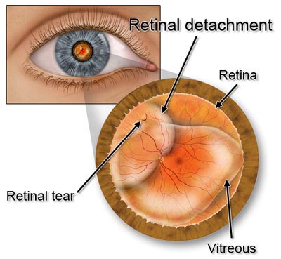 The retina is a layer of nerves at the back of your eye that senses light and then sends images to your brain. Your eye is like a camera. The lens in front of your eye focuses the light to the retina. In a way, you can think of the retina as film that covers the back of the camera like wallpaper. It needs to stay flattened against the back of the eye to work properly.
Your eye is filled with a clear gel called vitreous. This gel is usually clear and smooth. As you age, however, it’s possible for the vitreous to pull away from its attachment to the retina at the back of the eye. It’s also possible for pockets or gaps to develop in the gel. Most of the time, this happens without causing any problems.
When It Goes Bad
Sometimes, though, the vitreous can pull and tear the retina in one or even multiple places. The vitreous can then pass through the tear, causing the retina to peel off the back wall of the eye just like wallpaper would. A detached retina can occur at any age, but it is more common at middle age or later.
When your retina pulls away from its normal position in your eye, it’s called a retinal detachment. The retina doesn’t work when it’s out of place in your eye. Retinal detachment can turn into a very serious problem that almost always causes blindness if not treated quickly.
If you are having any visual abnormalities you should always be evaluated with a thorough consultation and examination by a physician for an accurate diagnosis and treatment plan as it may be a symptom or sign of a serious illness or condition.
Conditions Make Retinal Detachment More Likely
There isn’t any one thing you can do to prevent the detachment of your retina. But regular eye exams can help to catch the symptoms early. And if you’re at a higher risk, you should make those exams an annual priority. Conditions that often lead to detached retina include:
- Nearsightedness
- Glaucoma
- Previous cataract surgery
- Serious eye injury
- Weak areas in the retina that can be seen in an eye exam
- Previous history of retinal detachment
- Family history of retinal detachment
- Health problems such as sickle cell disease or diabetes
Symptoms to Watch for
Your eyes are not something to take for granted. Instead, you need to report symptoms as soon as they occur to head off further consequences. Symptoms of a detached retina can include:
- A grey curtain moving in your line of vision
- A shadow on the edge of your line of vision
- Flashing lights or new floaters in the eye
- Cloudy, blurry, or wavy vision
These symptoms could indicate a detached retina, but symptoms aren’t always present. If any of these occur, call your ophthalmologist as soon as possible to either rule out or diagnose a detached retina.
You can also have holes or tears in your retina, which can happen with or without a detached retina. Holes or tears do not immediately affect your vision, but if left untreated, they can lead to a detached retina. This leads to serious vision problems and needs to be treated immediately.
If you are having any abnormal visual symptoms, you should always be evaluated with a thorough consultation and examination by a physician for an accurate diagnosis and treatment plan as it may be a symptom or sign of a serious illness or condition.
Obtaining a Diagnosis
Your eye doctor discusses your health history with you and performs an exam. The doctor does a number of tests to determine if there are any problems with your eye or the systems inside it. Some of the tests include:
- A “slit lamp” exam to look closely at the front and back of your eye
- An ultrasound exam, which uses sound waves to look at the back of your eye
- An exam using eye drops to dilate your pupils so that a light can be used to look at the back of your eye
Treatment
Your doctor can treat a retinal tear or hole by laser treatment or cryotherapy. This seals the holes to prevent more vitreous from leaking behind your retina. This treatment causes little to no discomfort and can be done in your ophthalmologist’s office. If done quickly enough after the injury, it can prevent your retina from detaching. Sometimes, retinal tears and holes heal by themselves without treatment.
A detached retina may require surgery to restore it to its correct position at the back of your eye. This can happen several different ways. The method used depends on the position of your retina, the severity of the detachment and any underlying conditions that may have contributed to the tear. Regardless of the surgical method used, retina doctors repair any tears or holes as part of the same surgery.
Surgical Methods
Surgery to repair a detached retina is called pneumatic retinopexy. This process involves injecting a gas bubble into the vitreous inside the eye. The gas bubble pushes the retina back against the eye wall where it belongs. Laser or cryosurgery is then used to repair any tears or holes and to anchor the retina in its proper position.
After the surgery, you’ll be asked to keep your head in a specific position for a few days to make sure the process is successful. The gas bubble itself dissipates over time as your eye heals. Your ophthalmologist can do this procedure in the office.
Other surgical procedures may include:
- Scleral Buckle, during which a flexible band or buckle is put around the center of your eye. This serves as a counterbalance for any force that could be pulling the retina out of its ideal position. Your doctor may also drain the vitreous fluid from behind your eye, which allows the retina to return to where it should be. This procedure is done in an operating room, but usually on an outpatient basis.
- Vitrectomy is a procedure done to drain the vitreous gel, to keep it from pulling on the retina. It may also need to be done if the vitreous is being replaced by a gas bubble. If this is the case, your body fluids eventually replace the vitreous, because the vitreous does not return once it is drained. On occasion, a vitrectomy is combined with a scleral buckle if necessary. This surgery is also done in an operating room.
After surgery, your vision may take several months to improve. It’s also possible that any vision lost due to the detached retina may not return. Allow yourself ample time for recovery, follow your eye doctor’s instructions closely and resume normal activities gradually.
If you are having any abnormal visual symptoms, you should always be evaluated with a thorough consultation and examination by a physician for an accurate diagnosis and treatment plan as it may be a symptom or sign of a serious illness or condition.
Important Reminder: This information is only intended to provide guidance, not a definitive medical advice. Please consult eye doctor about your specific condition. Only a trained, experienced board certified eye doctor can determine an accurate diagnosis and proper treatment.
Do you have any questions about retinal tear or retinal detachment surgery in NYC? Would like to schedule an appointment with the top rated Ophthalmologist specialist in NYC, Optometrist Dr. Saba Khodadadian of Manhattan Eye Specialists, please contact our office for consultation with eye doctor.
Manhattan Eye Specialists
Dr. Saba Khodadadian, Optometrist (NYC Eye Doctor)
Dr. Saba Khodadadian, Optometrist (NYC Eye Doctor)
983 Park Avenue, Ste 1D19
New York, NY 10028
New York, NY 10028
(Between Madison Ave & Park Ave)
☎ (212) 533-4821
DISCLAIMER: PLEASE READ CAREFULLY
The information on this website is to provide general guidance. In no way does any of the information provided reflect definitive medical advice and self diagnoses should not be made based on information obtained online. It is important to consult a best in class Optometrist or Ophthalmologist in NYC regarding ANY and ALL symptoms or signs as it may a sign of a serious illness or condition. A thorough consultation and examination with an eye specialist should ALWAYS be performed for an accurate diagnosis and treatment plan. Be sure to call your local eye doctor or call our office today and schedule a consultation.